Imaging Experts
OUR SERVICES
At Pueblo Radiology, we provide the newest and most advanced imaging services. Find specifics about each modality we specialize in including what to expect, frequently asked questions and appointment preps.
Osteoporosis Risk Factors
- Age: increased age, increased risk
- Gender: females greater than males
- Family history
- Race: Caucasian, Asian, greater than Hispanic, African-American
- Bodyweight: low body weight, increased risk.
- Menopause
- Lifestyle: cigarettes, excess alcohol, increased risk
- Medications: steroids, certain anti-seizure medications.
Who Should Undergo Bone Density Testing:
- All postmenopausal females, females with history of premature menopause.
- Patients with an unexplained fracture.
- Patients on medications which increase risk of osteoporosis such as steroids.
- Patients considering hormone replacement therapy or other treatment for osteoporosis.
- Osteoporosis on radiographs.
- History of osteoporosis, follow up of treatment for osteoporosis.
Prevention of Osteoporosis:
- Eat a balanced diet rich in calcium and Vitamin D.
- Take daily multivitamins and calcium supplements.
- Do weight-bearing exercise.
- Reduce smoking and alcohol intake.
- Hormone replacement therapy and medications may also be indicated. Discuss medical treatment options with your referring physician.
- Avoid unsafe conditions which can lead to falls and fractures.
- Discuss your medications and their effect on your bone density with your referring physician.
- Bone density testing is recommended to know where you stand as far as bone health.
BEFORE THE EXAM:
PREPARATION FOR THE EXAM
- There is no special preparation. You may eat normally.
- There is no need to disrobe if you wear clothes without buttons, zippers or other metals. Loose fitting clothes such as workout outfits, sweatpants, and sweatshirts are recommended.
- You should not have this exam if you have had any kind of barium study within the last week.
- You will be asked to fill out a questionnaire prior to your scan to help your radiologist in interpreting your bone density test.
WHAT TO EXPECT
The test will take approximately 15-20 minutes. You will be lying on your back throughout your exam. The actual scanning of the spine, hips and wrists only takes a few minutes. The exam is painless with no injections. Your exam will be reviewed by our radiologist who specializes in bone density interpretations and results will be sent to your referring physician.
Ultrasound
Vein Treatment
Screening / Preventative
01.
CT Cardiac Calcium Scoring
02.
CT Lung Screening (Low Dose)
03.
Screening Vascular Ultrasounds
CT Cardiac Calcium Scoring
Vascular disease is among the leading causes of death in the United States, yet is generally asymptomatic until a catastrophic event occurs, such as a heart attack, stroke or aneurysm rupture. Pueblo Radiology offers preventive vascular screening ultrasound to bring legitimacy and medical integrity to the “parking lot” screening exams commonly seen in many communities.
Pueblo Radiology’s vascular screening covers three important areas:
- Carotid arteries (neck) – to access stroke risk
- Aorta (abdomen) – to detect the presence of aortic aneurysm
- Blood pressure assessment of the lower extremities to identify PAD (1) and risk of heart disease
Together, this three-part assessment can be an important element in determining risk of vascular disease in general — implying relative risk in areas that are more difficult to “see”, such as the heart (coronary arteries).
Individuals 55 years of age or older with any of the following cardiovascular risk factors may benefit from preventative screening for vascular disease:
- Diabetes mellitus
- History of hypertension
- Hypercholesterolemia
- Known cardiovascular disease
- Smoking
Noninvasive screening examinations have proven to be accurate in detecting vascular disease prior to active warning signs and before a major medical incident such as stroke or sudden death from aneurysm rupture. With the baby boomer population aging and Americans 60 years old expected to reach nearly 76 million by 2020, raising public awareness about vascular disease and screening becomes critical.
Pain Management
At Our Office, The Following Procedures Are Performed:
The procedures below require an order from your physician.
Epidural Spine Injection
An epidural steroid injection (ESI) is the delivery of powerful anti-inflammatory medicine directly into the space outside of the sac of fluid around your spinal cord. This area is called the epidural space.
Transforamenal Injection (Spine)
Epidural glucocorticoid injections are commonly given to patients with leg and/or back pain to relieve such pain and improve mobility without surgery.
Facet Joint Injection (Spine)
A facet joint injection is performed to treat neck and back pain in combination with other non-surgical spine treatments like rest, medications, chiropractic manipulations, and physical therapy.
Joint Injections
Joint injections are non-surgical procedures that apply a solution of anesthetic and corticosteroid directly to damaged joint tissue.
Tendon Injections
In a tendon injection, your foot and ankle orthopaedic surgeon puts a substance into either your tendon or the area around your tendon with a needle.
Bursa Injections (Around Major
Joints)
Bursa injections contain steroids that soothe bursitis inflammation and joint pain. The steroid injection eases symptoms of hip bursitis, shoulder bursitis and other types of bursitis
PUEBLO RADIOLOGY’S INTERVENTIONAL SPECIALISTS

Interventional Procedures
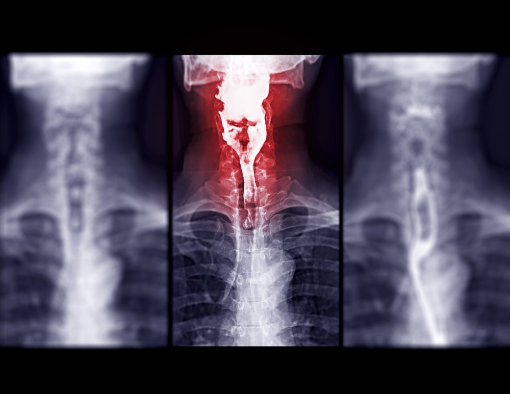
CT Scan
What are x-rays?
Since their discovery in 1895 by the German physicist Wilhelm Roentgen, X-rays have played a major role in helping physicians diagnose and treat disease. X-rays are high-energy electro-magnetic waves created within an x-ray tube.
They are highly penetrating, and in combination with computer imaging plates, provide images of various internal organs and structures.
Your physician and the radiologist combine to provide you with the test best suited to your particular situation. While all radiation exposure carries some risk, the benefits of diagnosing your condition far outweigh these risks.
However, due to these risks, X-ray examinations are carried out by trained, licensed personnel and interpreted by physicians (radiologists) who are specially trained in the imaging sciences.
Pueblo radiology x-ray services:
1. GI Studies (Barium)
Barium is an X-ray absorber and appears white on X-ray film. When swallowed, a barium drink coats the inside walls of the upper GI tract organs so that the swallowing motion, inside wall lining, function, size, and shape of these organs are visible on X-ray.
2. Hysterosalpingography
Hysterosalpingography is a test to determine whether a woman’s Fallopian tubes are open, as well as if there is any disease in her uterus.
3. Urinary Tract Studies
An IVP is a study of the kidneys and bladder and the tubes connecting them called the ureters.
GI Studies (Barium)
The prep for an UGI is found in our appointment prep section. During your exam, the radiologist uses a TV-like x-ray device (fluoroscope) to watch the barium travel down your esophagus (“food pipe”) and into your stomach and small bowel (intestine).
The radiologist will take pictures of the various structures of your GI tract as he/she instructs you to turn from side-to-side.
FAQs
You belong to a high-risk group if any of the following factors apply to you:
- You have high cholesterol levels
- You have high triglyceride values
- You smoke
- You have high blood pressure
- You are hereditarily at risk
- You have diabetes
Or any of the factors in combination with a sedentary lifestyle. If one or more of the above points apply to you, contact your physician to find out more about how CT can help in the evaluation of your heart.
Mammography
at santa barbara women’s imaging center
Mammography and all and other breast imaging and breast procedures are offered at our sister facility.
Click the logo below to be redirected to Santa Barbara Women’s Imaging Center to learn more, set up an appointment or contact us.
MRI & Whole body mri
magnetic resonance imaging
What Is Magnetic
Resonance Imaging?
Magnetic Resonance Imaging (MRI) is a sophisticated medical imaging
technique that uses powerful magnets and radio waves to generate detailed images of the inside of the body. Unlike X-ray or CT scans, MRI does not use ionizing radiation, making it a safe and noninvasive option for medical
diagnosis. MRI is particularly useful for visualizing soft tissues and frequently used when evaluating organs, muscles, joints, breast tissue, the spine, and the brain. Its versatility and ability to produce detailed images make MRI an essential tool in modern medicine for accurate diagnosis and treatment
planning.
click for more info +
what to expect:
BEFORE
Prior to your exam, you may continue your normal dietary regime. Upon your arrival, we will ask you to change into a cotton gown and remove all metal objects if you have not already done so. Some of these objects include jewelry, metal zippers or buttons, hairpins and hearing aids. Please notify our office ahead of time if you have any metal devices inside your body or if you are pregnant.
DURING
For the duration of your exam you will be asked to lie still on a table so that the MRI can produce the highest quality images. During scanning, there will be a loud (at times very loud) knocking noise; during imaging, we will provide you with earplugs and/or headphones with music to minimize this noise. You will also be able to communicate through an intercom with our technologists if you are feeling any discomfort. The entire exam usually lasts around 20 to 30 minutes; some exams can last up to one hour.
AFTER
After your exam, you can return to your normal routine. Our highly trained radiologists will analyze your images and report the findings to your doctor.
OUR TECHNOLOGY
PHILIPS MR5300 1.5T
In the first quarter of 2024, Pueblo Radiology invested in a new MRI technology from Philips Medical designed for those patients with claustrophobia or MRI-based anxiety. The Ambient Experience unique to this scanner, provides the patient with an immersive viewing experience for scans involving the head, neck, upper extremities, chest, and abdomen. Soothing communication and music will be heard through headphones, keeping the patient informed as they move through their scan. As with our other MRI scanners, this new scanner also has a large opening with a short bore design, meaning that for most of the lower extremity exams, the patient’s head will be out of the scanner. Lastly, the scanner’s advanced technology will provide faster scan times, meaning less time in the scanner for the patient. As compared to “open” MRI scanners, our scanner is faster with significantly better image quality.
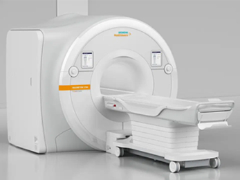
siemens verio 3t
Our Verio magnet has a large opening, 27 1/2”, the same as our 1.5T MRI scanner. In addition, this machine accommodates patients of up to 550lbs. With the strength of the magnet, Pueblo is able to acquire outstanding images with relatively short exam times.
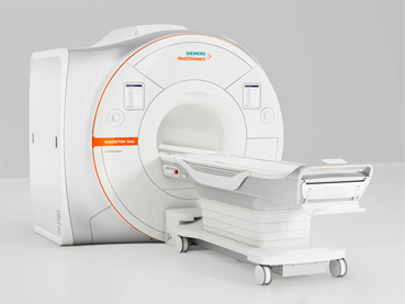
SIEMENS ALTEA 1.5T
In September of 2021, Pueblo Radiology upgraded one of our existing MRI scanners. The new platform called Magnetom Altea is a 1.5T MRI scanner with features that support state-of-art imaging as well as maximum patient comfort. The user-friendly operating system, combined with a sophisticated scanning capability, allows us to reduce the time patients spend in on the scanner. The magnet itself has a patient-friendly space with a wide aperture and short bore length, combined with soothing lighting and air conditioning within the scanner. Patients having scans on older scanners have been impressed with the shortened exam times and high level of comfort provided by this new system.
FAQs
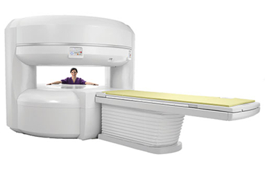
Pueblo also supports an “open” MRI unit which does not involve the tunnel associated with MRI. The open unit is open on the sides and can be seen here on our website. Pueblo Radiology has the largest open bore (“closed”) scanner in the region. The 70 cm high field system generates extraordinary images. Some patients require the use of a true open scanner. We are fortunate to provide the option of an open bore scanner for those patients. Our Open MRI office is located at 31 West Gutierrez Street in downtown Santa Barbara.